Molluscum Contagiosum

By Courtney McClurkin, DPM; Andrew Olson, DPM

Molluscum contagiosum can be confused with a variety of different skin pathologies

History

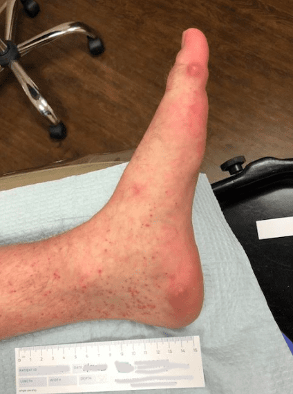
A 25 year old male with no known significant past medical history presents with a pigmented lesion to plantar aspect of his left foot present for an undetermined period of time.

Physical Examination

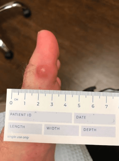
A raised red and black 3mm lesion is seen on the plantar aspect of left foot.

Diagnostic Testing

A 2mm punch biopsy was performed with clinical concern to rule out melanoma.

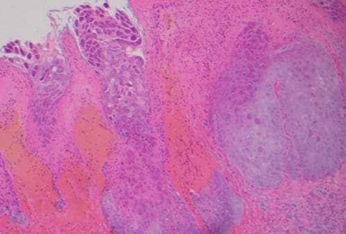
Histological Findings

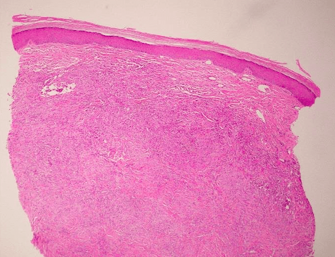
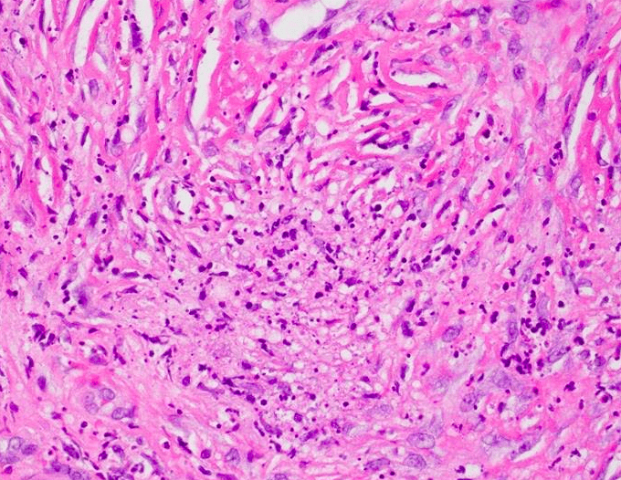
Typical histological features of molluscum contagiosum were identified including molluscum bodies, which are intracytoplasmic eosinophilic structures contained within epidermal cells.  These molluscum bodies become basophilic in the upper part of the epidermis, and are found in high numbers in the horny layer.  There is also a dense polymorphonuclear infiltrate noted adjacent to the epithelium containing the molluscum bodies.

Click on image to enlarge

Click on image to enlarge

Diagnosis

Molluscum contagiosum

Discussion

Molluscum contagiosum is a skin infection that is caused by a poxvirus.  Transmission of the molluscum contagiosum virus is through contact with an infected person or contaminated surface.  It is most commonly found in young children, but is also notorious for sexual transmission among adults and adolescents.  Transmission rates are generally similar for the immunocompromised.  However, the lesions can be more numerous, appear in atypical locations, and be more resistant to treatment.  The virus incubates for a period of 14 days to 6 months, followed by formation of slowly enlarging, single or multiple lesions.  The lesions most commonly appear as pearly, umbilicated papules.  They can become inflamed and erythematous, causing a “molluscum dermatitis”.  Most cases are self-limiting, though crops of lesions may come and go over the span of months.  There are, however, a plethora of treatment options ranging from physical destruction to systemic treatments.

Clinically, molluscum contagiosum can be confused with a variety of different skin pathologies, including but not limited to: verruca vulgaris, condyloma, nevocellular nevus, basal cell carcinoma, keratoacanthoma, lichen planus, eczema, foreign body granuloma, sarcoidosis, folliculitis, and true epidermal cyst.

Two histologic variants exist.  The pseudocystic variant is distinguished by invagination of colonized epithelium of dilated horn-filled infundibula bordered by a thick epidermis and are filled with compact, thick keratin.  In the polyploid variant, the dermis located beneath molluscum contagiosum has loose collagen fibers, dilated vessels, and inflammatory infiltrate.  Molluscum bodies are the most distinguishing  histologic feature.  These are large cells with cytoplasmic eosinophilic inclusions that contain viral particles.

Associated lesions with molluscum contagiosum are common, including true epidermal cysts, nevocellular nevus, Unna nevus, Clark nevus, soft fibromas, and Kaposi sarcoma.  Abscesses can be associated due to discharge of molluscum bodies to the dermis, followed by release of proinflammatory cytokines and activation of complement pathway.

Treatment

While molluscum contagiosum is typically self-limiting within 6 months, there are treatment options available.  Treatment is initiated in order to alleviate discomfort produced by itchy or painful lesions, for cosmetic reasons, to prevent spread of lesions, as well as to prevent trauma, scarring, and secondary infection of lesions.

Surgical treatments include: cryotherapy, curettage, punch biopsy, manual excision, electric cauterization, and laser therapy.

Topical treatments include: acidified nitrite, adapalene, Australian lemon myrtle oil, benzoyl peroxide, bromogeramine, cantharidin, cidofovir, diphencyprone, griseofulvin, honey, hydrogen peroxide, imiquimod, iodine, phenol, podophylotoxin (HIV patients), potassium hydroxide, salicylic acid, tea tree oil, and tretinoin.

Systemic treatments include cimetidine, griseofulvin, and cidofovir (in HIV-infected patients).

References:

Cribier, B., Y. Scrivener, and E. Grosshans. Molluscum contagiosum: histologic patterns and associated lesions. The American Journal of Dermatopathology 23(2): 99-103 (2001).

Mancini, A.J. and A. Shani-Adir. Other Viral Diseases. Dermatology 3rd ed. (2012): p. 1358-59.

Van der Wouden, J.C., et. al. Interventions for cutaneous molluscum contagiosum (review). Cochrane Database of Systematic Reviews Issue 5, Art. No.: CD004767 (2017).


Erythema Elevatum Diutinum

By Desiree Dalcherone, DPM; Laura Fernandez, DPM; Pooja Srivastava, DPM

Lesions are usually asymptomatic, but some cases might present with itching and pain

History

Patient is a 29 year old male with past medical history of Crohn’s disease, presenting with a one year history of progressively worsening, painful nodules to his bilateral legs and feet.

A punch biopsy of skin was taken from the nodule present to the patient’s plantar-medial left hallux.

Physical Examination

Multiple nodules were present on bilateral legs and feet.

Click on image to enlarge

Click on image to enlarge

Click on image to enlarge

Histologic Findings

Sections show an intense neutrophilic infiltrate with associated nuclear dust and interstitial eosinophils at all levels of the reticular dermis. The deeper regions of the reticular dermis show significant fibrosis in association with the infiltrate present. Active neutrophilic vasculitis is felt to signify a late stage process.  Gomori methenamine silver stain fails to demonstrate fungal elements.  AFB stain fails to demonstrate mycobacterial organisms.

Diagnosis

Erythema Elevatum Diutinum (EED)

Discussion

Erythema elevatum diutinum is categorized as a chronic fibrosing leukocytoclastic vasculitis usually occurring in patients in their thirties to fifties, earlier in HIV. The lesions might present as orange to yellow plaques, or red to purple papules and nodules measuring approximately up to 1 cm. They are usually found in extensor surfaces in the back of the hands or fingers, wrists, elbows, knees, ankles, and toes. The lesions are usually asymptomatic, but some cases might present with itching and pain, usually made worse with cold environments.

Inflammatory bowel disease (Crohn’s disease and ulcerative colitis) has been associated with the onset of erythema elevatum diutinum.  EED has also been reported in patients with Celiac disease and resolved with a gluten-free diet.

Other reported associations include rheumatoid arthritis, granulomatosis with polyangiitis, relapsing polychondritis, pyoderma gangrenosum, Sweet syndrome, cutaneous lupus erythematosus, nodular scleritis and panuveitis, Hashimoto thyroiditis, juvenile idiopathic arthritis, Sjögren syndrome, dermatomyositis.  EED can be associated with systemic infections, including streptococcal infections, HIV, hepatitis B, tuberculosis, and syphilis

ANCAs (60% IgA and 33% IgG) are found in EED, however, other ANCA-positive vasculitides such as granulomatosis with polyangiitis and microscopic polyangiitis, do not present with these lesions.

Differential diagnosis includes granuloma faciale which also involves the dermis. Granuloma faciale, however, shows mostly eosinophils whereas EED shoes more neutrophils. Sweet Syndrome is another form of neutrophilic disease with rapid onset of tender and erythematous plaques and nodules, but it is differentiated by the lack of leukocytoclastic vasculitis.

Treatment

Since EED is generally associated with an underlying systemic disease, such as hematologic conditions, autoimmune disease, chronic infections, HIV- it is imperative to control the underlying cause as well as directly targeting the lesions.

In this particular case, the patient’s EED is likely associated to Crohn’s Disease. Treatment for Crohn’s Disease include anti-inflammatory drugs such as aminosalicyclates, corticosteroids, sulfasalazine and azathioprine. Aminosalicyclates are generally first-line treatment, and suppress inflammation to the intestine. Antibiotics such as tetracyclines and sulfa drugs can also be given adjunctively to kill bacteria in certain areas of the bowel, and also decrease inflammation.

Focusing on the nodules themselves, oral Dapsone is the most common drug given, with a response rate of up to 80%. Dapsone works by inhibiting neutrophil chemotaxis. Second-line drug treatments include corticosteroids, colchicine, and methotrexate.

Surgical excision of the nodules in a progressed disease state has also been found to provide symptomatic relief, however since EED nodules are quite recurrent, multiple surgeries are needed to keep the patient nodule-free.

References:

Rinard, J., R. Mahabir, J. Greene, and P. Grothaus. Successful surgical treatment of advanced erythema elevatum diutinum. Can J Plast Surg 18(1): 28-30 (2010).

Wollina, U., C. Kronert, A. Koch, J. Schonlebe, A. Vojvodic, T. Lotti. Erythema elevatum diutinum – two case reports, two different clinical presentations, and a short literature review. Global dermatology. 18(7):3039-3042 (2019).

Crohn’s Disease. Harvard Health Publishing : Harvard Medical School. https://www.health.harvard.edu/a_to_z/crohns-disease-a-to-z. November 2018.

Calonje E, McKee PH, undefined undefined undefined. Vascular Diseases. In: McKees Pathology of the Skin: with Clinical Correlations; 5th ed. Philadelphia, PA: Elsevier, Saunders; 2020:714-770

James WD, Elston DM, Treat JR, Andrews GC, undefined undefined undefined. In: Andrews Diseases of the Skin: Clinical Dermatology. 13th ed. Elsevier; 2020:813-861

Sandhu JK, Albrecht J, Agnihotri G, Tsoukas MM. Erythema elevatum et diutinum as a systemic disease. Clinics in Dermatology. 2019; 37(6): 679-683.