How Epidermal Nerve Fiber Density is used to diagnose and treat patients with small fiber peripheral neuropathy

Diagnostic confirmation allows for the most effective treatment 

BakoDx’s epidermal nerve fiber density (ENFD) analysis is an objective diagnostic tool used to monitor and manage patients with small fiber peripheral neuropathy (SFPN).

The analysis not only provides definitive confirmation of SFPN, but also enables better assessment of the severity of the neuropathic process, which may have treatment implications. ENFD analysis may also reveal degenerative changes in those patients with normal densities, which may predict the future onset of SFPN.

SFPN may present with varying symptoms, however, most commonly presents with burning/shooting pains, pins and needle sensation or numbness and coldness over the extremity. The neuropathic process typically progresses in a distal to proximal fashion, or in other words, in a “stocking and glove” type distribution. These symptoms are in contrast to those seen in large fiber peripheral neuropathy (LFPN) in that tendon reflexes and muscle strength are typically normal, as well as NCV/EMG are also within normal limits. While there are “textbook” symptoms one would look for in SFPN, this is often not as specific as one would like. Due to the varying presentations associated with SFPN, as well as cross over symptoms with large fiber peripheral neuropathy, diagnostic confirmation is essential. Once diagnostic confirmation is acquired, clinical identification of the underlying etiology, if possible, will allow for the most effective treatment. In many cases an etiology can be determined, with Diabetes (type I and II) being the most common and others including autoimmune disorders, inherited, or infectious causes. However, up to 40 percent of SFPN cases are idiopathic in nature.

The sample submitted for ENFD analysis is a simple, quick, minimally-invasive, 3mm punch biopsy procedure of the distal leg (10cm proximal to the lateral malleolus). The procedure is performed in an office setting, limiting outside referral. The biopsy itself requires approximately five minutes, with little to no post-procedure care and both the procedure and the test are reimbursable by nearly all payers.

The ENFD analysis requires a specially trained pathologist to count and calculate the nerve fiber density of the myelinated A-delta and unmyelinated C fibers within the epidermis. The direct observation of these small intra-epidermal nerve fibers makes ENFD the most objective method for diagnosing SFPN.

The therapy for SFPN is usually directed towards the underlying disease process or symptom relief. Therapies could include diet/lifestyle modifications, NeuRx-TF or other medical food products, topical anesthetics, and central acting agents to alleviate symptoms.

For more information, including procedural videos, ordering a complimentary ENFD kit, or request an in-service, visit bakodx.com/enfd.


Why ENFD Analysis Provides the Best Tool to Confirm Small Fiber Peripheral Neuropathy

One quick in-offi­ce procedure can provide a definitive diagnosis, allowing for effective treatment and symptom relief

Have patients complained of burning pain, tingling, numbness, or a pins and needles feeling of the lower extremity?

This could be an indication of small fiber peripheral neuropathy[1] (SFPN), a condition that can be definitively confirmed with a quick, in-office punch biopsy procedure and epidermal nerve fiber density (ENFD) analysis.

“For the early detection of small fiber peripheral neuropathy, ENFD analysis is the best objective tool,” says Dr. Wayne L. Bakotic, Medical Director at BakoDx. “The ENFD diagnostic test allows for the confirmation of SFPN, the assessment of the degree and severity, and determination of effective treatment.”

During ENFD analysis, a specially trained pathologist calculates the density per unit area and quantifies the tiny myelinated A-delta and unmyelinated C fibers within the epidermis. This highly specific and sensitive testing method allows clinicians to definitively diagnose suspected SFPN.

This in-office procedure is a minimally invasive, 3mm punch biopsy of the distal leg (10cm proximal to the lateral malleolus). It typically takes about five minutes, with little to no post-procedure care. The ENFD test is reimbursable by most insurance companies.

Watch Video

Neuropathy is complex

While peripheral neuropathy affects about 20 million adults in the U.S.[2], it is often misdiagnosed with large fiber neuropathy due to its complex array of symptoms, both somatic and autonomic. Patients with SFPN will often have normal reflexes, muscle strength and electromyography (EMG) and nerve conduction velocity (NCV) test results.

SFPN usually affects the hands and/or feet in a stocking or glove-like pattern.[3] A patient may present with symptoms that range from burning and tingling, to coolness and numbness. This form of neuropathy often occurs secondary to diseases such as diabetes mellitus or it may be an indication of other autoimmune disorders.

Other patient presentations may include:

  • Restless leg syndrome
  • Abnormalities in sweating (hyperhidrosis)
  • Inability to sweat normally (anhidrosis)

But because these presentations are not always SFPN, identifying the precise form of neuropathy allows for the most effective treatment method.

ENFD test benefits

ENFD is a powerful tool that provides the most objective method of documenting and monitoring the progression of SFPN. Early ENFD testing may also reveal degenerative changes, so that patients are placed on preventative therapies prior to the onset of symptoms.

Once the diagnosis of SFPN is confirmed, there are several medical treatments that may provide relief, depending on the type of nerve damage. Therapy to treat the symptoms and underlying causes of the neuropathy could include diet/lifestyle modifications, supplements, or topical medications. However, because SFPN has many causes, no product works for everyone.

Studies show that dietary supplements, such as NeuRx-TF, containing Alpha-Lipoic Acid[4] (600mg daily) Benfotiamine[5] (600mg daily), may be helpful in some cases to diminish symptoms of neuropathy and improve epidermal nerve health. Investigators also have noted a benefit to using combination therapy that includes L-methylfolate, methylcobalamin and pyridoxal 5’-phosphate[6].

ENFD retesting at future intervals, between six-to-12 months, may also help to assess the disease progression or regression and the therapy effectiveness over time. An accurate and definitive diagnosis of SFPN provides improved patient care and allows for appropriate neuropathy management.

“My patients have seen a drastic improvement in their quality of life through providing ENFD analysis and subsequent treatment,” said Lilly Khavari, DPM, a Texas-based physician-owner. “Your patients will love you for offering this test.”

[1] Hovaguimian A, Gibbons CH. Diagnosis and treatment of pain in small-fiber neuropathy. Curr Pain Headache Rep. 2011 Jun;15(3):193-200. doi: 10.1007/s11916-011-0181-7. PMID: 21286866; PMCID: PMC3086960.

[2] Peripheral Neuropathy Fact Sheet | National Institute of Neurological Disorders and Stroke. Ninds.nih.gov. https://www.ninds.nih.gov/Disorders/Patient-Caregiver-Education/Fact-Sheets/Peripheral-Neuropathy-Fact-Sheet. Published 2020. Accessed August 20, 2020.

[3] Cascio MA, Mukhdomi T. Small Fiber Neuropathy. In: StatPearls. Treasure Island (FL): StatPearls Publishing; May 23, 2022.

[4] Ziegler D, Low PA, Litchy WJ, et al. Efficacy and safety of antioxidant treatment with α-lipoic acid over 4 years in diabetic polyneuropathy: the NATHAN 1 trial. Diabetes Care. 2011;34(9):2054-2060. doi:10.2337/dc11-0503

[5] Luong KV, Nguyen LT. The impact of thiamine treatment in the diabetes mellitus. J Clin Med Res. 2012;4(3):153-160. doi:10.4021/jocmr890w

[6] Walker MJ Jr, Morris LM, Cheng D. Improvement of cutaneous sensitivity in diabetic peripheral neuropathy with combination L-methylfolate, methylcobalamin, and pyridoxal 5′-phosphate. Rev Neurol Dis. 2010;7(4):132-139.


Current Concepts in Psoriasis Therapy

Largely unchanged for decades, treatment methods for this potentially disabling condition are finally evolving.

Introduction
Our ability to treat adverse medical conditions has long been an admixture of miraculous accomplishment and frustrating stagnation. For every disease such as osteogenic sarcoma, where recent therapeutic breakthroughs have lifted cure rates from 10-20% to over 90%, there is a condition like pulmonary adenocarcinoma, where cure rates approach their historic controls. The management of dermatologic disorders has not offered any particular exception to this rule. There recently have been profound advances in the medical management of inflammatory conditions of the skin; however, many of those advances are the first since the advent of artificial corticosteroids several decades ago. Moreover, an effective means of medically managing some of the most aggressive neoplastic processes of skin remain elusive. For instance, the prognosis attributed to melanoma and Merkle cell tumor, once they’ve spread beyond their site of origin (stage 3 or 4), remains exceedingly poor.
The management of psoriasis has remained largely unchanged for decades. With a better understanding of the pathogenesis of this potentially disabling condition, investigators have begun targeting the precise factors that lead to its progression. This new focus has shown extraordinary promise for long term medical management, particularly in severe cases. In this report we will discuss the clinical findings, diagnosis, and evolving methods of treatment for psoriasis with special attention given to those cases that present themselves in the lower extremity.

The precise inciting event in the development of psoriasis remains unclear.

Epidemiology
The overall prevalence of psoriasis approaches 2 percent internationally; however, it has been estimated to be as high as 4.6% in the United States.1 The prevalence in children is significantly lower, calculated at 0.5% in a large series of kids ages 12-16.2 The incidence in persons of color is lower than that in Caucasians, affecting roughly 0.7 percent of Africans and Asians.1 There is no particular gender predilection.1 The epidemiological features of palmoplantar (acral) psoriasis has remained somewhat obscure; however, in our experience, neither palmoplantar pustulosis nor the chronic plaque form of acral psoriasis can be considered rare in a podiatric practice, and both are certainly under-diagnosed. There is a much rarer form of pustular psoriasis which is limited to the digits and/or nails. This rare variant is designated “acrodermatitis continua”.

There are two peak ages for the development of psoriasis, the first being during early adulthood and the second being during the sixth decade of life.3 Cases manifesting in childhood are far more likely to be associated with HLA-Cw6 and a positive family history.4,5 Such patients also trend toward more severe forms of psoriasis with associated arthritis and nail involvement.6

Pathogenesis
The pathogenesis of psoriasis has been much studied and fairly well characterized; however, the precise inciting event in its initial development remains unclear.7 Psoriasis is fundamentally an autoimmune disease whereby CD4-positive, and to a lesser extent CD8-positive, T lymphocytes are stimulated to evoke a type-1 inflammatory response. Central to this response is the liberation of pro-inflammatory cytokines such as TNF-alpha which have the net result of producing local tissue damage and increasing the turnover rate of the epidermis. Where normal squamous epithelial cells require roughly 15 days to traverse from the basal layer to the stratum corneum, in affected epithelium, this rate is reduced to 1-3 days.8 This rapid turnover of affected squamous epithelium results in the silver scale that is characteristic of psoriasis. Due to the rapidity with which affected epidermis turns over, there is insufficient time for the formation of a normal stratum corneum. In some instances there may be a genetic component to the development of psoriasis. Its inheritance is in the form of a polygenic trait, resulting in the development of psoriasis in 14% of offspring when 1 parent has the disease, and 41% of offspring if both parents are affected.9 There are associations with various major histocompatibility complex phenotypes, particularly HLA B13, HLA B17, and HLA Cw6. The presence of HLA Cw6 antigens confers a relative risk of 13 for the development of psoriasis in the Caucasian population.10 A major gene involved in the development of psoriasis has recently been mapped to chromosome 6p21.3 and has been designated as PSORS1.11 Psoriasis may come and go without apparent reason; however, there are also numerous potential triggers. In many instances, psoriasis is distributed upon sites that are most affected by friction or pressure. This is the result of Koebner phenomenon, a term used to denote conditions that may be catalyzed by external trauma. Other triggers include alcohol ingestion, HIV infection, streptococcal pharyngitis (guttate variant of psoriasis), certain drugs (lithium, glucocorticoids, and beta-blockers), and emotional stress.12,13 Emotional stress plays a particularly important role as a trigger in children.12

Acral psoriasis is limited to the volar surfaces of the hands and/or feet in 81% of cases.

Clinical Findings
In the most common form of psoriasis, well-delineated geographic plaques form upon sites that are predisposed to external trauma. This common form of psoriasis has been aptly designated as the chronic plaque form of psoriasis vulgaris. When following its classic pattern of distribution, the knees, elbows, sacral region, and scalp are affected. Focal involvement of the skin is not uncommon even in the chronic plaque form of psoriasis, where approximately 2/3’s of patients develop a limited and relatively mild expression of the disease.14
Chronic plaque form psoriasis is characteristically covered by “silver” scales which when removed produced small areas of pin-point bleeding (Auspitz sign). Such silver scales are less prominent on the palms and soles. When the volar surfaces of the hands and/or feet are involved, the condition is referred to as acral psoriasis. This form of psoriasis breaks with the pattern of distribution exhibited by routine psoriasis vulgaris. Acral psoriasis is unique in that it is limited to the volar surfaces of the hands and/or feet in 81% of cases.9 It is patients with this form of psoriasis that most commonly present themselves to podiatric clinicians. There are many potential nail manifestations in psoriasis. Such pathologic changes vary from the classically described pitting and “oil spots” to onycholysis and frank keratinizing nail dystrophy. Though one set of investigators estimated the frequency of nail involvement to be roughly 80%, the true rate of involvement is probably slightly lower.15 Such overestimation is probable because bona fide keratinizing psoriatic nail unit dystrophy may be clinically indistinguishable from onychomycosis, idiopathic onycholysis, and traumatically induced dystrophy. In the lower extremity, keratinizing nail unit dystrophy (resembling onychomycosis) (Figure 1)

Figure 1: Psoriatic nail involvement most commonly presents as a keratinizing nail unit dystrophy (Courtesy Sean VanMarter, DPM). Figure 1: Psoriatic nail involvement most commonly presents as a keratinizing nail unit dystrophy (Courtesy Sean VanMarter, DPM).

is much more likely to be identified than is pitting of the nail plate or “oil spots” (Figure 2).16

Figure 2: “Oil pits” may be seen in the lower extremity; however, they more commonly manifest on the fingers (courtesy Andrew Levy, DPM). Figure 2: “Oil pits” may be seen in the lower extremity; however, they more commonly manifest on the fingers (courtesy Andrew Levy, DPM).

In addition to the chronic plaque-forming type of psoriasis, there are more eruptive or acute forms of psoriasis, descriptively designated as pustular and guttate (drop-like) psoriasis. Although these forms of psoriasis may precede or co-exist with chronic plaque psoriasis, due to their unique clinical presentations, they are by convention considered separately. The topic of acral psoriasis warrants separate discussion because of its tendency toward unusual clinical presentations, its recalcitrance to first line therapeutic modalities, and its common role as a diagnostic pitfall. Acral psoriasis includes those cases of psoriasis that predominantly involve the volar (non-hair-bearing) surfaces of the hands and/or feet. As aforementioned, in the overwhelming majority of cases, persons with this variant of psoriasis lack skin involvement beyond the acral surfaces. While chronic plaque form psoriasis may affect the feet in its classic form (Figure 3a and 3b),

Figure 3a: Chronic plaque form psoriasis may affect the feet in its classic form, particularly on the dorsal surfaces (courtesy Brenna Steinberg, DPM). Figure 3a: Chronic plaque form psoriasis may affect the feet in its classic form, particularly on the dorsal surfaces (courtesy Brenna Steinberg, DPM).

Figure 3b: Occasionally plantar lesions may also demonstrate the classic silver scales (courtesy Don Heilala, DPM). Figure 3b: Occasionally plantar lesions may also demonstrate the classic silver scales (courtesy Don Heilala, DPM).

acral plaques are often less likely to demonstrate the characteristic silver scales that are typical of chronic plaque psoriasis when it arises in non-acral sites (Figure 4a and 4b).

Figure 4a

Figure 4b Figure 4b
Figure 4a and 4b: Plaques in acral psoriasis are less likely to demonstrate silver scales characteristic of chronic plaque psoriasis (courtesy J Percival, DPM).

In some cases, acral psoriasis presents as a diffuse keratoderma with thick gray keratotic plaques (Figure 5).

Figure 5 Figure 5: In some cases, acral psoriasis presents as a diffuse keratodermawith thick gray keratotic plaques (courtesy M.Gagnon,DPM).

Alternatively, acral plaques may be poorly demarcated with erythematous fine scale (Figure 6),

Figure 6: Acral plaques may be poorly demarcated with erythema and a fine scale (courtesy BenWeaver, DPM). Figure 6: Acral plaques may be poorly demarcated with erythema and a fine scale (courtesy BenWeaver, DPM).

or might present as round coinshaped lesions that are reminiscent of nummular dermatitis (Figure 7).

Figure 7: Acral psoriasis may present as coin-shaped lesions reminiscent of nummular (eczematous) dermatitis (courtesy Barry Blass, DPM). Figure 7: Acral psoriasis may present as coin-shaped lesions reminiscent of nummular (eczematous) dermatitis (courtesy Barry Blass, DPM).

Regarding distribution, some cases may not only involve, but are also limited to, the forefeet (Figure 8).

Figure 8: Some cases of acral psoriasis may be limited to the forefeet (courtesy Bruce Theall, DPM). Figure 8: Some cases of acral psoriasis may be limited to the forefeet (courtesy Bruce Theall, DPM).

In many instances, psoriasis will consist of only one or a few discrete plaques, while in others it involves the entire plantar surface. There may, or may not, be associated nail involvement and such nail involvement may be seen in isolation (Figure 9).

Figure 9: Psoriatic nail involvementmay be seen in isolation. Figure 9: Psoriatic nail involvementmay be seen in isolation.

The differential diagnosis is limited when chronic plaque psoriasis vulgaris presents in its classic distribution; however, because the acral variant may lack many of the distinguishing features for psoriasis (characteristic distribution, silver scale, well-demarcated plaques, Auspitz sign), it may be confused with a wide variety of dermatological conditions. Focal presentation of the chronic plaque form of psoriasis may be confused with conditions as disparate as eczematous (nummular) dermatitis, chronic allergic contact dermatitis, lichen simplex chronicus, or squamous cell carcinoma in-situ (Bowen’s disease). Acral psoriasis may also be mistaken for chronic T. rubrum dermatophytosis, acral lichen planus, pityriasis rubra pilaris, mycosis fungoides, or chronic dyshidrotic dermatitis. Finally, the pustular form of acral pustular psoriasis is commonly misdiagnosed as tinea pedis, resulting in the institution of errant anti-fungal treatment regimens for extended periods of time.

Diagnosis
There is no diagnostic dilemma when psoriasis presents with all its classic clinical features. Well demarcated plaques with silver scale over extensor surfaces may be considered psoriasis until proven otherwise.
Other helpful clinical clues to the diagnosis of psoriasis are Auspitz sign (pinpoint bleeds upon the removal of silver scales) and Koebner phenomenon (psoriatic lesions following the path of external trauma). Unfortunately,
acral psoriasis usually lacks the classic topographic distribution that is so often attributed to chronic plaque psoriasis. In addition, acral plaques often fail to exhibit the clinical features that allow them to be easily classified as psoriatic in nature. A potentially helpful sign, particularly when dealing with acral lesions, is the presence of
individual lesions in various stages of development, e.g., the presence of acute lesions (pustules), subacute
lesions (resolving pustules), and chronic plaques in the same individual.

When the diagnosis of psoriasis is in doubt, biopsies may be indicated. The technique of choice for the diagnosis of psoriasis and virtually all other inflammatory conditions of skin is punch biopsy. On the shoe-covered skin of the foot, many clinicians prefer to perform two 2mm punches biopsies rather than 1 large punch. This technique
allows for more rapid wound healing, better sampling, and reduced risk of infection. From a histopathologic perspective, performing two small punches allows for better sampling of the lesion in question. Sampling surface keratin alone will allow clinicians to rule out dermatophytosis, and may provide histopathologic features that are sugges tive of psoriasis; however, it rarely provides for a definitive diagnosis.

Treatment
Since the dawn of modern medicine, breakthroughs in the medical management of psoriasis have been few and far between. Advancements were delayed for centuries because of widespread confusion between psoriasis vulgaris and leprosy. Psoriatic patients were often expelled from society along with persons harboring Mycobacteria leprae infections in an errant attempt to ward off disease transmission. It wasn’t until the nineteenth century that these conditions were distinguished from each other, thereby allowing the development of effective therapeutic modalities for psoriasis. The oldest legitimate treatment for psoriasis was sunlight (ultra violet light). Many subsequent attempts at therapy were developed and subsequently abandoned during the 19th, 20th and early 21st centuries. As late as 1925 many purely nonsensical treatments were advocated for the treatment of psoriasis; amongst them: intramuscular mercury or bacterial antigen injections, non-protein diets, tonsil removal, and tooth extractions! 17 Despite the frightening nature of some of these early therapeutic approaches, others such as salicylic acid, coal tar, and ultra violet light (UVB) remain in use today.
When designing a treatment regimen for an affected patient, several factors must be taken into account. The clinical extent of involvement, the patient’s perception of that involvement, the subtype of psoriasis, the impact that some therapeutic modalities will have on lifestyle, and the potential side effects that medication might have.
The lower extremities may be affected by either non-acral chronic plaque form psoriasis, or acral (palmoplantar)
psoriasis. For the clinician, the significance of subtype relates to the potential for anatomic progression, the degree to which the condition will be debilitating, and its tendency toward recalcitrance. Although acral psoriasis
(cases limited to the non-hair-bearing surfaces of the sole and digits) may remain limited with regard to its anatomic scope, this variant may be extremely disabling and recalcitrant to traditional first line therapeutic
approaches. Chronic plaque form psoriasis is often less disabling, though more widespread, and tends to be more responsive to first line therapy.

Topical Corticosteroids
As a general rule, when less than 5% of the body surface area is affected by newly diagnosed psoriasis, the first line therapy should be limited to topical modalities. The possible exception would be for cases where the condition has already proven debilitating. For decades, the first line treatment of choice for limited psoriasis has been topical corticosteroids.18

Steroids of medium to suprahighpotency (Stoughten-Cornell classification 4, 5, 6, 7) are most commonly used for limited lower extremity psoriasis, either alone, or in combination with additional topical agents. Although there is technically a potential for iatrogenic Cushings syndrome after extended courses, the expression of such is rare and typically minimal. The same is true for the development of hypothalamic-pituitaryadrenal axis suppression and significant cases are usually limited to small children due to their low relative body surface area.19 Side effects such as skin atrophy and fragility are much more common complications of topical steroid therapy. Similar to other potential complications, clinically significant cases usually require long term use; in addition, those effects are most prominent on the steroid-sensitive skin of the face and intertriginous areas.18 Newer products such as fluticasone propionate have demonstrated great promise in further reducing the risk of adverse side effects. 20 Tachyphylaxis (decreased effectiveness with continued use) is a common problem with topical high potency corticosteroids; however, its presentation may be delayed or diminished by defaulting to a pulse-dosing regimen after an initial 2-3 course of b.i.d. applications. Such pulse-dosing may be accomplished by limiting applications to 3 application times over a 24 hour period once weekly.21 An alternate method of pulse dosing involves twice daily application for two consecutive days followed by 5 days off. In addition to the emergence of improved corticosteroid formulations, new vehicles such as gels and foams are gaining broad acceptance.

Vitamin D3 Analogues
Vitamin D3 analogues were introduced into the market for the treatment of psoriasis relatively recently, but have quickly joined corticosteroids as a first line therapy for the treatment of limited psoriasis. Within affected skin, vitamin D3 analogues inhibit epidermal proliferation, promote normal stratum corneum development, and inhibit neutrophil and monocyte/ histiocyte activity.22 Possibly the best known and most studied vitamin D3 analogue is calcipotriol  (Dovonex); however, other examples include calcitriol (Silkis) and tacalcitol (Curatoderem). Topical calcipotriol has been shown to be effective for the treatment of limited chronic plaque form psoriasis, including the palmoplantar variant. The efficacy of the vitamin D3 analogues is roughly comparable to that of a class 2 corticosteroid such as betamethasone dipropionate.23
Dosing for calcipotriol (50ug/g) is twice daily; however, night-time application twice weekly under occlusion has been shown to be equally effective in the treatment of palmoplantar psoriasis.24 The most significant clinical improvement is usually seen in the initial 6-8 weeks after initiating therapy.

Corticosteroid—Vitamin D
Analogue Combinations
Recently, formulations combining high potency corticosteroids and vitamin D analogues have been shown to be of clinical benefit. One such formulation combines betamethasone dipropionate and calcipotriol (Dovobet®). Such products are typically applied topically once daily and their long-term use has been shown to have a high safety
profile (see below).

Topical Retinoids
Tazarotene is a topical retinoid that was recently approved for the treatment of psoriasis. This product is available in 0.05% and 0.1% concentrations with a gel or cream base. Similar to vitamin D analogues, tazarotene lacks the side effects that are associated with long term corticosteroid therapy; however, this product has its own local side effects, in particular, irritation at the treatment site. This irritation may be managed by using combination therapy with a topical corticosteroid (see below). Due to the combination of its efficacy and its local side effects, tazarotene is usually considered a form of second line therapy in the management of limited psoriasis.
Roughly half the patients using the 0.05% or 0.1% gel describe a 50% improvement in their symptoms
after 6 weeks.25 Tazarotene gel has proven more effective than fluocinonide 0.05% cream.26

Combination Regimens Using
Topical Corticosteroids
As previously alluded to, corticosteroids are not simply effective as a form of monotherapy; they are widely used in combination with, or as an adjunct to, other topical medications. When used in combination with a keratolytic agent such as 5-10% salicylic acid or 40-50% urea, the therapeutic effects of the steroid are potentiated. In addition, corticosteroids decrease irritation that may be caused by salicylic acid. Corticosteroids also calm the local irritation that might be seen in association with retinoids and vitamin D3 analogues. In one study, improved efficacy and decreased irritation was noted when combining either tazarotene with a class IV (mometasone furoate 0.1% cream) or class II (fluocinonide 0.05% cream).27 The application of tazarotene 0.1% gel on Mondays, Wednesdays, and Fridays in alternation with clobetasol propionate ointment (class I) on Tuesdays and Thursdays, has led to the remission of psoriasis in 73% of patients with psoriasis.28 The combination of calcipotriol (vitamin D3 analogue) and betamethasone dipropionate (class I corticosteroid) has also been shown to be safe and effective.29

Ultra Violet Light and PUVA
Ultra violet light may be used in the treatment of psoriasis as either ultraviolet B phototherapy or photochemotherapy using UVA in combination with systemic or topical psoralen. Soaking affected feet in baths containing 8-methoxypsoralen followed by immediate UVA irradiation has shown to be particularly beneficial for the treatment of palmoplantar psoriasis while limiting side effects.30 The use of a topical cream containing 8- methoxypsoralen followed by UVA irradiation has proven equally effective in at least one small series.31 A recent publication noted the effectiveness of light emitted from a xenon-chloride excimer laser (308 nm) in the management of palmoplantar psoriasis.32

Systemic Therapeutic
Modalities
Many of the traditional forms of systemic therapy for severe psoriasis have remained unchanged—namely, Methotrexate and Cyclosporine remain staples in the management of patients with extensive disease. One new class of drugs that has shown notable promise for the management of psoriasis has been designated as the “biologics”. The biologic agents act at the cellular level to target specific processes that are involved in the development of a particular disease. Because their mechanisms of action are specific they tend not to have the widespread side effects that are associated with more globally acting agents such as corticosteroids. The
three principle forms of biologics are monoclonal antibodies, fusion proteins, and recombinant cytokines or growth factors.33
The generic names of the monoclonal antibodies and fusion proteins have been standardized to reflect their mechanism of action. For instance, the generic names of monoclonal antibodies have the suffix -mab, and receptor–antibody fusion proteins have the suffixcept.34 Biologic agents have one or more of 4 principle mechanisms of action. They 1) reduce the number of pathogenic T-cells (alefacept), 2) inhibit T-cell activation or migration (efalizumab), 3) modulate the immune system (Ilodecakin), or 4)
block the activity of pro-inflammatory cytokines such as tumor necrosis factor (etanercept).34

The prototypical T-cell reducing agent is alefacept, which, as its name implies, is a protein fusion product combining a T-cell binding site (Lymphocyte Function Antigen- 3) and sequences from the constant region of IgG’s heavy chain. This agent functions by binding with CD2, which is an antigen that is found on the surface of T-cell lymphocytes, particularly memory effector T-cells.35 After binding, this agent induces cell death through
apoptosis.35 Current dosing regimens call for consecutive weekly intramuscular injections of 15mg alefacept
for 12 weeks followed by 12 weeks of rest. At the conclusion of the rest period, a second course may be considered. A baseline CD4+ lymphocyte count should be performed prior to the initiation of therapy and then biweekly thereafter. In one study, 57% of patients experienced at least a 50% reduction in the Psoriasis Area Severity Index (PASI). This agent has also shown to provide a durable remission. Seventy four percent of those
patients that experience >50% reduction in PASI, maintain that such for the 12 week rest period.35

The second strategy for biologic agents used in the treatment of psoriasis involves the inhibition of Tcell
activation and migration. Efalizumab is an antibody that is directed against T-cell surface antigen CD11a (a subunit of Lymphocyte Function Antigen-1). Lymphocyte function antigen-1 (LFA-1) functions in T-cell activation and T-cell adhesion to keratinocytes and vascular endothelium.36 By interfering with the function of LFA-1, efalizumab inhibits both T-cell activation and the migration of T-cells into affected skin. The treatment regimen for efalizumab consists of the subcutaneous injection of 0.7mg/kg for the initial dose followed by 1mg/kg per week thereafter.
Higher dosage regimens and extended treatment periods are currently under  investigation. Initial results suggest added benefit with successive treatment cycles and higher dosages of 2mg/kg.37 The effectiveness of efalizumab appears to be slightly shy of that achieved by alefacept with a similar tendency toward durable remissions. The third mechanism by which biologic agents may influence the development and/or progression of psoriasis is through immune system modulation. Immune system modulation may be achieved by guiding the differentiation of T-cells toward forms that play a less pathogenic role in the development of psoriasis. In general, lymphocytes may follow one of two paths of differentiation. Lymphocytes that are CD4+ may differentiate into either Th1 or
Th2 phenotype and CD8+ lymphocytes may differentiate into either Tc1 or Tc2 phenotype. Lymphocytes
may be pushed toward the Th1 and Tc1 phenotypes through the influence of cytokines Il-12 and INF-gamma or toward the Th2 and Tc2 phenotypes through the modulatory effects of IL-4, IL-6, and IL-10. Psoriasis has been shown to be driven by the cytokines that are liberated by Th1 and Tc1 phenotypes.38 Chief amongst those instigating cytokines are IFN-gamma, IL- 2, and TNF-alpha.38
Interleukin-10 is a type 2 cytokine which is under-expressed in psoriatic patients. This substance actively
opposes the type 1 cytokine response; therefore it could conceivably curtail the progression of psoriasis by reducing the liberation of cytokines IFN-gamma, IL-2, and TNFalpha. Subcutaneous injections of recombinant human IL-10 (Ilodecakin) have been shown to prolong remissions when administered in combination with other products39;
however, the effectiveness of this cytokine as a stand-alone therapeutic agent in the treatment of psoriasis has thus far been disappointing. 40 Oprelvekin is a human recombinant IL-11 that has shown initial promise when administered daily in subcutaneous injections.41 This cytokine also diminishes the type 1 cytokine response; however, it further acts by decreasing both intraepidermal T-cells and keratinocyte adhesion molecules that facilitate the action of T-cells on affected epidermis. 41 Studies are currently ongoing to further assess the usefulness of these agents.The final, and possibly most promising, class of biologics for the treatment of psoriasis includes those that directly or indirectly block the activity of type-1 inflammatory cytokines. A chief target of this class of biologics is tumor necrosis factor-alpha (TNF-alpha). Tumor necrosis factor-alpha is a type-1 cytokine that functions as a potent mediator of inflammation and keratinocyte hyperproliferation. This factor plays a pivotal role in the development and escalation of psoriasis due to its excess production and liberation by T-cell lymphocytes, keratinocytes, and mast cells.42 In theory, reducing the activity of this cytokine should impede the pathogenesis of psoriasis. Etanercept (Enbrel) is a recombinant TNF-alpha receptor which is fused with the fc portion of IgG1. This product binds with TNF-alpha on cell membranes, thereby neutralizing it. This drug is administered weekly by subcutaneous injection in 25mg or 50mg doses. Investigators have shown that higher dosages and longer therapeutic regimens provide further benefit.43 In one study nearly 50% of all patients experienced either “complete clearing” or “almost complete clearing” of their lesions after 12 weeks of biweekly 50mg injections compared to 5% in the placebo group.44 An added benefit of etanercept therapy is the durability of disease remissions in those that respond favorably. The median time until relapse (<50% PASI) was 85 days with 25% of patients not experiencing relapse for 141 days.
Infliximab (Remicade) is an additional agent that inhibits the function of TNF-alpha; however, unlike etenercept, infliximab is a monoclonal antibody. Infliximab is a composite human-mouse antibody that targets TNF-alpha directly by binding it on the surface of cells. This agent is administered intravenously at 2-4 week intervals in a concentration of 5mg/kg. As many as 80% of patients with moderate to severe psoriasis can expect to have a 75% reduction in PASI and most will experience such benefits within 4 weeks.45
As a group, the biologics are extremely new agents and thus their safety profiles remain in flux. Injection site complications are the most common adverse effects associated with etanercept and tend to diminish with ongoing therapy.46 Another potential complication is an increased risk of infection, particularly upper respiratory infections. Chronic fatigue, liver toxicity, leukocytopenia and lymphopenia are rarely reported complications.46

Summary
Psoriasis is a chronic autoimmune condition of skin which may have unconventional features when arising on the acral surfaces of the hands and/or feet. Due to its less specific appearance when arising on the skin of the feet, misdiagnoses are common, potentially resulting in inappropriate treatment. First line therapy in the treatment of limited psoriasis remains topical corticosteroids; however, there is increasing use of Vitamin D3 analogues in this context. Combination therapy using both corticosteroids and Vitamin D3 analogues or corticosteroids and retinoids have been proven affective as a second line therapy. Advances in our understanding of the athogenesis of psoriasis has allowed for the introduction of a new class of effective therapeutic agents designated as the “biologics”. These medications exert their effects by directly inhibiting the function or production of the cytokines that are responsible for the development and propagation of psoriasis. Further long-term studies are needed; however, in initial research the biologics have demonstrated tremendous promise for the treatment of moderate to severe psoriasis, including debilitating variants such as that which affects the acral surfaces.

Largely unchanged for decades, treatment methods for this potentially

disabling condition are finally evolving.

Normal 0 false false false IN X-NONE AR-SA /* Style Definitions */ table.MsoNormalTable {mso-style-name:"Table Normal"; mso-tstyle-rowband-size:0; mso-tstyle-colband-size:0; mso-style-noshow:yes; mso-style-priority:99; mso-style-qformat:yes; mso-style-parent:""; mso-padding-alt:0cm 5.4pt 0cm 5.4pt; mso-para-margin-top:0cm; mso-para-margin-right:0cm; mso-para-margin-bottom:10.0pt; mso-para-margin-left:0cm; line-height:115%; mso-pagination:widow-orphan; font-size:11.0pt; mso-ascii- mso-ascii-theme- mso-hansi- mso-hansi-theme- mso-bidi- mso-bidi-theme-}


Needle Aspiration Biopsy

Needle aspiration biopsy is amongst the most underutilized diagnostic procedures of those available. This technique may be effectively used to rule out high‐grade malignancies when faced with nonspecific subcutaneous masses, in particular, those masses that resemble ganglion cysts. I once had three different medico‐legal cases under consultative review at one time, all of which were centered on patients who had non‐specific ganglion‐like masses. In each instance, the patient actually harbored a high‐grade sarcoma, and in each case, they were followed into their grave by their clinician with the errant diagnosis of “ganglion”. It is not overtly surprising that neoplasms masquerading as ganglion cysts may fool clinicians. For instance, roughly 70% of all the soft tissue masses in the foot are ganglia. This may lull clinicians into complacency, believing that all hoof sounds are derived from horses, and that zebras don’t exist. In 1999, Scully et al. of  Duke University summarized their experience with synovial sarcoma primary to the foot. In their series of 14 cases, 8 patients were followed for extended periods of time with the incorrect diagnosis of ganglion cyst. In our series of 401 pedal soft tissue tumors assembled at Memorial Sloan‐Kettering Cancer Center we had 8 synovial sarcomas. Amongst these 8 cases, 2 patients saw their iagnosis dramatically delayed because of the errant diagnosis of “ganglion”. Needle aspiration differs somewhat from most other biopsy techniques in that it provides the pathologist with cells and tiny tissue fragments to review, rather than large pieces of tissue. In other words, pathologists are not able to review a lesions overall architecture and pattern of growth. Instead, they must extrapolate the necessary diagnostic data from the appearance of individual cells. Because the material at the  athologists’disposal may be somewhatlimited, cytopathology can be somewhat less specific than histopathology. In this vein, the pathology reports derived from aspiration specimens typically provide basic, though highly significant information such as: “malignant cells not identified”, “atypical cells identified”, or “malignant cells identified”. Though vague in comparison to the diagnoses rendered after histopathologic analysis, these techniques may provide invaluable information in the management of patients with non‐specific subcutaneous masses, by ruling out the presence of high‐grade malignancies. The purpose of needle aspiration biopsy is to harvest cells and small pieces of tissue from lesions in question. To accomplish this, clinicians should use large (18 gauge) needles, and syringes that will produce high vacuum pressure (10cc or larger). An anesthetic wheal may be raised at the needle entry site. The needle is placed in the mass percutaneously, and the plunger is drawn back to create a vacuum, which is maintained through the procedure. The needle is partially withdrawn, and then redirected into each quadrant while maintaining the vacuum. Once each quadrant has been sampled, the vacuum is released, and the needle is removed. If fluid is obtained, it may be put directly into fixative. If no aspirate is apparent, fixative should be drawn up into the syringe, and then the collective contents is returned to the specimen jar. In this context, the ICD‐9 is 238.1 and the CPT code is 10021 (10022 when performed with imaging guidance).


Cutaneous Biopsy Techniques in the Management of Chronic Wounds

Many clinicians rely exclusively on clinical acumen when determining how to manage chronic wounds. Though an ulcer’s clinical features may be fairly indicative of its etiology, in some instances, such is not the case. Even among the most characteristic‐appearing ulcerations, masqueraders do exist. Ruling out the possibility of an unsuspected neoplastic or inflammatory condition could be necessary for the

successful management of chronic wounds. In this context, cutaneous biopsy techniques may be invaluable; however, their utility does not necessarily end here. There are three common clinical settings in which a biopsy may be used in the management of a chronic wound. Clinicians may use histopathology to 1) confirm a clinically suspected diagnosis at the outset of care, to 2) rule out a mimic in cases where a wound is showing recalcitrance or unusual progression, to 3) assess for an underlying predisposing condition independent of the ulceration, or to 4) assess for compounding feature, such as an excessive bacterial burden. Because the clinical presentation of cutaneous  ulcerations may be virtually pathognomonic of a particular etiology, the first of these scenarios should not always give rise to a biopsy; however, in some instances, confirmation is warranted. In a minority of cases, the clinical manifestations that surround an ulceration are entirely nonspecific and a biopsy is indicated prior to the initiation of medical care. For wounds that appear characteristic of a particular etiology, biopsies are usually not initially necessary; however, as a rule of thumb, biopsies should be considered for all ulcers that cannot be readily explained or fail to show improvement after 2 months of treatment. In instances such as this, biopsies are being used to verify that the implemented therapeutic regimen is appropriate. Delays in the diagnosis of some mimics may be medicolegally treacherous. For instance, malignant melanoma, particularly amelanotic variants, may create ulcers that are virtually identical to non‐neoplastic ulcers. Delays in this diagnosis may have serious implications with regard to the affected patient’s outcome. Simply stated, the failure to reassess ones differential diagnosis in cases where ulcerations show unusual clinical behavior, or recalcitrance, may be a direct cause of increased morbidity. An additional clinical setting where a biopsy might prove useful in the management of chronic ounds, involves patients with suspected neuropathy as a predisposing condition. With a 3mm punch biopsy of skin, taken for 10 cm above the lateral malleolus, physicians may qualify and quantify the presence of small fiber neuropathy. Degenerative changes among the intra‐epidermal nerves, further may be predictive of the future onset of small fiber neuropathy. Though this examination uses a simple 3mm punch of skin, there are differences in the handling of biopsies taken for this purpose. Most important among these differences are that punches taken for epidermal nerve fiber density testing require a specialized fixative that must be requested from the lab, and care must be taken to avoid crushing the surface epithelium when removing the tissue from the biopsy site. Formalin fixative renders the biopsies useless for small fiber analysis. In most instances, the biopsy technique of choice for verifying the cause of an ulceration, assessing for neoplastic and non‐neoplastic mimics, and characterizing predisposing conditions, is a punch biopsy. In the initial two settings, a central and peripheral 3mm punch is usually


Biopsy Planning and Post-Biopsy Tissue Handling : A Synopsis

<!--

brad-bakotic

-->

Introduction

As a general rule, the podiatic profession is notorious for its underutilization of cutaneous and soft tissue biopsy techniques. In some instances, such under-use may be attributed to trepidation stemming from a lack of familiarity with the biopsy techniques themselves; however, I believe this is not the preeminent cause. In my experience, the most common reason that many podiatric clinicians don’t use biopsy techniques to their fullest is a degree of insecurity regarding when to use witch biopsy and what to do with them once the biopsy has been performed. In most offices, the support staff is ill prepared to offer much in the way of guidance, leaving the clinicians on their own.

The purpose of this report is to summarize the indication for biopsy, when each technique is most appropriate, and the method of fixation that is required for laboratory examination.

As a general rule, the podiatric profession is notorious for its under-utilization of cutaneous and soft tissue biopsy techniques.

Biopsy Techniques

Not uncommonly, clinicians create unnecessary limitations with respect to how they should use biopsies and what constitutes a ‘biopsy’ itself. We can easily fall into a pattern of thinking whereby we equate the word ‘biopsy’ with a 3:1 ellipse or limit the word biopsy to only those specimens taken by punch technique. Either misimpression might result in under-utilization and the latter would likely result in suboptimal sampling in more than half of the cases in which a biopsy is being used. Simply stated, anytime tissue is taken to obtain a diagnosis through histopathologic analysis, a biopsy has been performed. Common techniques include, but are not limited to, punch biopsy, shave biopsy, saucerization (a form of shave biopsy), curettage, core needle, and aspiration biopsy.

Punch biopsy (CPT 11100 initial, CPT 11101 subsequent) is a common technique used to sample conditions that 1) are too large to be shaven or 2) have a deep dermal component (requiring deeper sampling). This is the ideal technique for sampling all forms of dermatitis; however, this is far from where its utility ends. Punches may be used to sample suspected vasculitis, ulcers, large pigmented lesions (those too large to be shaven), and other large suspected neoplasms (those that are greater than 1cm in diameter). Three-millimeter punches are used for epidermal nerve fiber density testing (Figure 1).

Figure 1 A 2mm punch biopsy. This technique is used for epidermal nerve fiber density testing, however a 3mm punch in necessary in that context (courtesy Carl Solomon, DPM). 
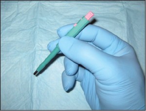
Figure 1. A 2mm punch biopsy. This technique is used for epidermal nerve fiber density testing, however a 3mm punch in necessary in that context (courtesy Carl Solomon, DPM).

Small punches (2mm) also may be ideal for plantar verrucous lesions. Because this technique samples the full thickness of such plantar lesions, it allows dermatopathologists to accurately discriminate between genuine verrucae and clinically identical verrucous carcinomas. In sum, punch biopsies are ideal when a small part of a much larger lesion is submitted for histopathology.

Standard shave biopsies (CPT 11300 series) are used much more commonly in dermatologic practices than are punches. In fact, the combination of standard shaves and saucerizations) represents the overwhelming majority of biopsies performed by dermatologists. This technique uses a scalpel to “shave off” a lesion of concern (Figure 2),

Figure 2 Standard shave biopsy. This is the procedure of choice for sampling papules and small nodules. 
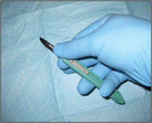
Figure 2. Standard shave biopsy. This is the procedure of choice for sampling papules and small nodules.

leaving a shallow iatrogenic ulceration in its wake. Shave biopsies contrast sharply with punch biopsies in many respects. Shaves do not routinely sample the deep dermis, rather, they typically extend only to the depth of the superficial dermis. In addition, where punches are relatively narrow (most punches are 2-4mm in diameter), shaves often encompass a broad sample of superficial skin (often 1cm or greater in width).

Shave technique is the ideal sampling method for unexplained papules (elevated lesions measuring 5mm or less in diameter), and can be used for macules (flat lesions measuring less than 1cm in diameter). Usually a standard shave is performed when the lesion in question appears superficial and/or exophytic (outward growing).

Clinicians create unnecessary limitations with respect to how they should use biopsies and what constitutes a ‘biopsy’ itself.

Saucerization (CPT 11300 series) is a technique that is closely related to a standard shave biopsy. In fact, saucerization has many of the same indications and is coded identically. This technique uses a bendable blade, not dissimilar to a Gillette razor blade, to “scoop out” the tissue of concern (Figure 3).

Figure 3 Saucerization biopsy. This technique is ideal when sampling flat lesions and those that require additional depth. 
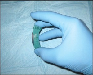
Figure 3. Saucerization biopsy. This technique is ideal when sampling flat lesions and those that require additional depth.

Where standard shaves are ideal for superficial and/or exophytic lesions, saucerizations are better for flat or endophytic (inward growing) lesions. Conceptually, this technique is thought of as a more aggressive method of sampling and thus can lend itself more to complete excision than can shave.

Curettage (CPT 11100 unless completely excised) is a less common sampling method than are the previously described techniques; however, it remains a viable biopsy technique for many dermatologists. Curettage uses a dermal curette to scrape the surface of the skin to obtain the desired tissue sample (Figure 4).

Figure 4 Curettage. This technique can be used for sampling superficial lesions which can be “scraped off” the skin surface. Figure 4. Curettage. This technique can be used for sampling superficial lesions which can be “scraped off” the skin surface.

The disadvantage of curettage is largely related to how difficult these specimens may be to histopathologically examine. Inherent in the mechanics of this technique is the fragmentation of the sample. This may make the tissue impossible to correctly orient, which can complicate its histopathologic examination.

Core needle biopsy is an ideal method of characterizing more deeply seated masses. This technique is perfect for investigating tumors of the soft tissue. Essentially any mass that falls into the differential diagnosis with a lipoma or ganglion could be sampled using c o r e n e e d l e technique. Makers of core needles include Temno ™ and Trucut™ (Figure 5).

Figure 5 Core needle biopsy. Pictured is a Temno™ core needle which is ideal for harvesting tissue from subcutaneous or deep soft tissuemasses. 
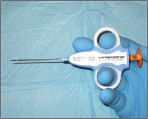
Figure 5. Core needle biopsy. Pictured is a Temno™ core needle which is ideal for harvesting tissue from subcutaneous or deep soft tissue masses.

Core needles a r e h o l l o w , large gauge needles designed to withdraw thin columns of tissue from within the mass in question. The technique is easy to perform and is highly effective. To obtain tissue using core needle biopsy, the skin overlying the mass is lanced and the instrument is inserted to abut the mass in question. Depending on the size of the mass, the biopsy instrument is set by the clinician to sample to a desired depth using a spring loaded mechanism. The device is discharged and the sample is obtained. This is usually repeated into the masses’ various quadrants.

Needle aspiration biopsy (CPT 10021 without imaging guidance or 10022 with imaging) is slightly less invasive than is core needle biopsy but has essentially the same indications. This technique uses a large gauge hypodermic needle and a large syringe to sample fluid and/or cells from deeply seated masses (Figure 6).

Figure 6 Needle aspiration biopsy. This is a useful technique to help guide the surgeon toward open biopsy, additional imaging, etc. 
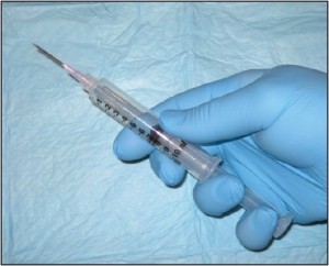
Figure 6. Needle aspiration biopsy. This is a useful technique to help guide the surgeon toward open biopsy, additional imaging, etc.

The secret of core needle biopsy is to create a vacuum once the needle has been introduced into the mass and to maintain the vacuum throughout the remaining aspiration procedure. The needle should be redirected into each of the 4 quadrants twice, without releasing the plunger and without completely removing the needle from the mass. Any fluid that is obtained may be expelled into fixative. If grossly perceivable tissue is not obtained, fixative should be drawn up into the syringe and then squirted back into the specimen container . This functions to flush any of the potentially neoplastic cells out of the aspiration needle.

Biopsy Indications The indications for biopsy vary depending on the nature of the lesion in question. As a general rule, there are three categories of indications for biopsy: 1) atypically appearing lesions, 2) typically appearing lesions which behave in an atypical fashion, 3) progressive, longstanding lesions or conditions which cannot be adequately explained. For inflammatory conditions / dermatitis, the indications are predominantly related to the patient’s perception of the condition. Indications include, but are not limited to: 1) significant patient frustration, 2) severe symptomatology, 3) recalcitrance and 4) those cases that have entirely nonspecific clinical findings. In contrast, the indications for biopsy in relation to pigmented lesions of the skin are almost entirely related to the clinician’s impression of the lesion in question. Asymmetry of pigment, asymmetry of configuration, irregular outlines, and a diameter of greater than 6mm all represent indications for biopsy.off” the skin surface.

Pigmented lesions include all those lesions that might fall within the differential diagnosis of a pigmented malignant melanoma. This differential diagnosis includes lesions as disparate as solar lentigos, melanocytic nevi, seborrheic keratoses, tinea nigra, and talon noir. The criteria which should lead to the consideration of biopsy within this class of lesions are: 1) asymmetry of configuration, 2) asymmetry or variegation of color, 3) irregular outlines, or 4) size greater than 6mm in diameter . The symmetry of configuration should be assessed in 3 dimensions. As such, if the lesion is bisected in a plane that is perpendicular to the skin, each half should be identical, not just with regard t o i t s s h a p e along the skin’s surface, but also its elevations and depressions. It should also be stated that newly arising or changing lesions greater than 6mm in diameter in adults represent an absolute indication for biopsy, not a relative one. The indications for biopsy in relation to non-pigmented tumors of the skin are less well defined and lend themselves to more subjectivity than those for pigmented lesions. For the most part , papules, nodules, and plaques that cannot be effectively explained should receive prompt biopsy, or at minimum, clinical follow-up. Lesions that do not show evidence of resolution or involution should be sampled. An important point here is that simply harboring a papule or nodule that cannot be explained is of itself “atypical”.

Frank evidence of malignancy is not ne c e s s a ry to prompt a biopsy. In keeping with this theme, biopsies should be considered on all ulcers that cannot be adequately explained, and those that persist despite appropriately targeted therapy. Additional miscellaneous biopsy indications include: verrucous lesions in persons over 40 years of age, tumors of all types that show recalcitrance, and pyogenic granulomata in persons over 40 (Figure 7).

Figure 7 This Kaposi’s sarcoma in a 40 yearold male could easily be confused with a pyogenic granuloma. (courtesy Carl Solomon, DPM). Figure 7. This Kaposi’s sarcoma in a 40 yearold male could easily be confused with a pyogenic granuloma. (courtesy Carl Solomon, DPM).

The soft tissue includes all the non-epithelial, non-skeletal, and non-visceral tissue of the body. This includes smooth muscle, skeletal muscle, adipose tissue, fibrous tissue, and peripheral neural tissue, amongst others. The indications related to biopsying tumors of the soft tissue are complicated by three facts. Foremost, tumors of the soft tissue are usually masked by the overlying skin and fascia, making their clinical features entirely nonspecific. Secondly, the foot boasts a large number of benign soft tissue masses that aren’t particularly common elsewhere within the body. Ganglia, for instance, are largely limited to the hands and feet. The same is true of digital mucous cysts. Finally, podiatric clinicians may be unaware of core needle and aspiration biopsy techniques, instead relying on more invasive open biopsies or complete excisions. This constellation of facts has the net result of diminishing the number of biopsies performed by podiatric clinicians and lowering their index of suspicion when dealing with soft tissue masses in the foot. In many instances, diagnostic procedures are delayed, resulting in tumor progression and a worsened prognosis. As a general rule, progressive soft tissue masses that cannot be adequately explained should be investigated. The dimension that is often given in the orthopedic oncology literature as an absolute indication for biopsy is 5cm; however, this is of little value when dealing with masses in the foot because of their easy accessibility. Tumors become evident at a much smaller size in the foot and thus should be investigated. Simply having a cystic component does not exclude the possibility of neoplasia as high grade sarcomas and many adnexal carcinomas can cavitate . Similarly, a mass’s tendency toward trans-illumination does not equate with benignancy .Many cystic and myxoid malignancies will trans-illuminate to some extent (Figure 8).

Figure 8 This high-grade fibrosarcoma was not biopsied because like a ganglion it transilluminated. Figure 8. This high-grade fibrosarcoma was not biopsied because like a ganglion it trans illuminated.

Imaging may be helpful; however, as many clinicians will acknowledge, this rarely provides a definitive opinion with regard to a mass’s potential biologic behavior. Early investigations may be aided by needle aspipapule ration biopsy. This technique has the advantage of being minimally invasive and well tolerated. This makes it an easy technique to perform as a purely investigative procedure, allowing clinicians to rule in or out the obvious diagnoses. Masses that fail to produce an aspirate(“dry taps”) should be taken for open biopsy, the most common form of which is core needle biopsy. When the aspiration of a mass produces fluid of an unusual consistency, it should be sent for cytopathologic analysis. Initial aspiration biopsies, which produce clear mucinous fluid typical of a ganglion, may be sent for analysis at the clinician’s discretion; however, subsequent aspirations performed on recalcitrant lesions must be analyzed.

Technique Selection

Large plaques, patches

The principles for technique selection are fairly rudimentary with few exceptions. Dermatitides and other conditions of the skin, which are too broad to “shave off”, are instead punched. Inflammatory conditions that would most often be punched include psoriasis, eczematous (nummular) dermatitis, lichen planus, granuloma annulare, and all conditions that might fall into their differential diagnosis. Neoplasms also may be punched in some instances. Squamous cell carcinoma in-situ may present as a plaque and thus warrant punch biopsy. Similarly,melanoma insitu may present as a broad patch or plaque that is more conducive to punch technique (Figure 9).

Figure 9 Melanoma in-situ may present as a large patch, which is best sampled with multiple punch biopsies (Courtesy Mark Lambert, DPM). 
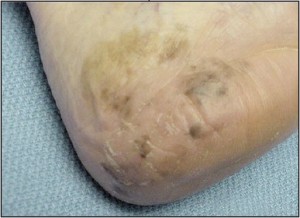
Figure 9. Melanoma in-situ may present as a large patch, which is best sampled with multiple punch biopsies (Courtesy Mark Lambert, DPM).

In general terms, when a small part of a patch or plaque is sampled for histopathology, punch technique is the technique of choice. One exception to the general rule that only large lesions are punched, and those that are small are shaven, involves verrucous lesions on the plantar surface. Because keratotic lesions iagof the plantar surface tend to be endophytic, shaves may not sample deep enough to obtain diagnostic tissue. For such lesions, punch technique will better allow the dermatopathologist to distinguish between lesions that may mimic each other superficially. An example of two lesions that might be identical superficially, and one that has caught me unsuspecting, are those of superficial palmoplantar wart and verrucous carcinoma.

Ulcers

Ulcers may be sampled by several methods depending largely on their clinical setting. Ulcers may be either neoplastic (carcinoma or melanoma) (Figure 10) or inflammatory (stasis or vasculitis) in etiology.

Figure 10 Neoplastic ulcers may closely resemble those that have medical etiologies.Such lesions may be easily sampled with a central and peripheral punch biopsy. 
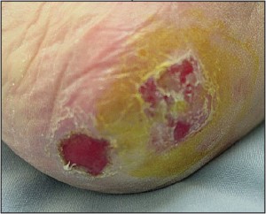
Figure 10. Neoplastic ulcers may closely resemble those that have medical etiologies.Such lesions may be easily sampled with a central and peripheral punch biopsy.

In most instances punches are ideal in either setting; however, exceptions exist. When punches are used, their ideal number and placement might vary. When characterizing suspected neoplastic ulcerations, a single punch may be all that is needed to obtain a diagnosis; however, in such conditions two are better than one to ensure appropriate sampling. In this clinical setting, one central punch and one upon the mounded flesh at the periphery of the ulcer is usually adequate. Usually relatively small punches are adequate. Alternatively, clinicians could potentially use a dermal curette or scalpel to obtain viable tissue from the ulcer’s base and proud peripheral flesh. In contrast to neoplastic ulcers, to characterize inflammatory ulcers, only punch biopsy techniques are recommended. In most cases, the cause of the ulceration in question lies deep within the dermis, far below the reaches of a superficial shave biopsy. For most conditions at least two punches are recommended, one central and one over the epidermal lip at the periphery of the ulcer. One exception to this recommendation would be for suspected vasculitis. In this context, multiple random punches are required over the ulcer base. In the case of vasculitis, the objective is to sample multiple locations along the ulcerbase in hopes of finding an affected vessel within the underlying dermis. Because this is a search that is reminiscent of depth charges seeking out an underlying submarine, there is an advantage to using punches of 3mm or larger.

Advanced superficial tumors

In most cases, the objective when biopsying advanced superficial tumors is not to rule in or out malignancy as much as it is to define the type of malignancy (Figure 11).

Figure 11 Advanced superficial neoplasms such as thismelanoma are sampled to precisely characterize a knownmalignancy. 
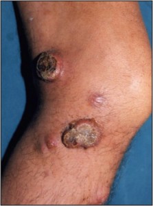
Figure 11. Advanced superficial neoplasms such as thismelanoma are sampled to precisely characterize a known malignancy.

This allows the clinicians to know whether they should deal with the neoplasm in question themselves, or whether the patient should be referred out to an alternative subspecialist. In addition, for those cases that are to be referred out, the precise diagnosis will provide the podiatric physician with the information s/he needs to refer the patient out to the appropriate discipline. In this setting, a punch biopsy will allow the clinician diagofnostic tissue without complicating a future excision or sentinel node biopsy.

Soft tissue/subcutaneous masses

Deep soft tissue masses and those lesions which clinically resemble them were discussed briefly above. To elaborate, there are four methods of sampling masses of the soft tissue: 1) excisional biopsy, 2) incisional open biopsy, 3) core needle biopsy, 4) needle aspiration biopsy. Excisional biopsy should be reserved for bases that are small and proven to be benign.

Shaves typically extend only to the depth of the superficial dermis.

The accidental excision of an unsuspected sarcoma could have prognostic significance ranging from loss of limb to loss of life. Incisional open biopsy carries with it the advantage that a larger volume of tissue is removed, which may be useful for testing purposes. However, due to the high risk of wound site complications, and poor tumor sampling, this technique is usually initially deferred. The most common technique for sampling soft tissue masses in the extremities is core needle biopsy. This technique allows the surgeon to harvest cores of tissue from various sites within the mass in question and has a very low complication rate. The most common core needles for this indication are Tru-cut™ and Tenmo™. Needle aspiration is an excellent initial minimally invasive technique to help verify benign diagnoses (ganglia), and to help guide clinicians to more invasive biopsy techniques when indicated.

Papules or macules

Papules and macules are by definition less than 1cm in greatest dimension (Figure 12).

Figure 12 Papules and macules such as this basal cell carcinoma are best sampled using shave technique or saucerization (Courtesy Mark Lambert, DPM). Figure 12. Papules and macules such as this basal cell carcinoma are best sampled using shave technique or saucerization (Courtesy Mark Lambert, DPM).

Because of their small size they can often be removed entirely, or almost entirely, using a standard shave technique or saucerization. By removing the preponderance of the lesion in question for histopathologic analysis, the dermatopathologist will be far less likely to be fooled by inadequate sampling. Papules are small elevations over the skin surface which measure less than 5mm (1cm in some texts). Because these primary lesions are elevated, they are readily shaved flush using a 15 or 10 blade. As papules become small nodules, they tend to become less superficial in extent. For relatively small nodules that seem to have a component that extends deeper into the dermis, or for macules (small flat alterations in pigment), saucerization is often a preferred biopsy technique. Because saucerization uses a metal blade that is bent to form an arch, this techniqueallows the physician to “scoop out” the biopsy site. The curve of the blade allows for better control when sampling lesions that lie deep to the skin surface.

Post-Biopsy Specimen Handling

The correct handing of the biopsy specimen, after the procedure has been performed, could make the difference between obtaining a rapid diagnosis and the performance of a wasted procedure. The overwhelming majority of tissue that is sampled using biopsy techniques should be fixed in formalin (10% formaldehyde); however, exceptions exist. The exceptions are as follows: Gout: When gout is suspected, the specimen should be placed in dehydrated alcohol. Alcohol preserves the monosodium urate crystals for crystal analysis and review under polarized light. Regardless of the biopsy method, alcohol is preferred unless the sample is kept fresh in a sterile container or a slide preparation is made in the office setting. Remember formalin contains 90% water, and water dissolves monosodiumu rate crystals.

Epidermal nerve fiber density testing: Formalin fixative is an excellent preservative for routine histopathology; however, it has deleterious effects on tissue taken for epidermal nerve fiber density analysis (degenerates intra-epidermal nerves and blocks important antibody binding sites on their surface). There are two potential fixatives that can be used for this test, each typically distributed only by labs specializing in this test (see www.bakopathology.com for more information).

Aspirations: Aspiration biopsies are ideally preserved in cytologic fixative. Formalin may dull some of the fine nuclear features that aid in diagnoses. Alternatively, slide preparations may be made in the office setting when clinicians have experience in this technique. We have made important diagnoses on aspirates that were received in formalin fixative; however, clinicians should know that this fixative has some limitations.

Cultures: Samples taken for microbiologic culture are not typically considered “biopsies” in the same sense as those procedures herein discussed and would not be billed in the same manner. Such samples are mentioned here only to stress that

tissue taken for culture should be kept fresh and not contaminated. This may be done using appropriate tissue swabs, agar slants, or sterile cups. There is a roughly 3 day window within which such specimens fixashould be plated after which bacterial cultures begin to be compromised. Formalin and alcohol will preserve tissue for histopathologic analysis, but it will kill all living cells within the sample.

The small size of most biopsies makes them particularly susceptible to air dry artifact

Most biopsies are small, either in width, depth, or both. Their small size makes them particularly susceptible to air dry artifact. Once samples that are to be analyzed histopathologically are placed in formalin fixative,they are indefinitely preserved. The case is similar with alcohol. Punch biopsies taken for epidermal nerve fiber density are unique in that they can be left in their special fixative (not formalin) for only 24 hours before the quality of the sample will begin to erode. For this reason, the specimen is typically retained within the clinical practice for 12-24 hours as it is being fixed, rinsed, and then placed in cryoprotectant for shipping.

Conclusion

In summary, there are a host of biopsy techniques within the armamentarium of today’s podiatric surgeon, each of which has its own indications. Many of the indications for these procedures are absolute and leave room for subjectivity only in unique circumstances. Once the indication has been acknowledged, the most appropriate biopsy technique should be chosen and performed. The product of most biopsies will be small ,making it necessary to quickly transfer the specimen into the correct fixasamtive. Formalin is usually the fixative of choice but exceptions do exist. Finally, although there is always an “ideal” technique for a particular indication, the fundamental principle is that “making the diagnosis trumps all else”. As physicians, we’ll virtually never do harm by doing a less than ideal procedure; however, in some instances, we will ensure harm by doing nothing at all.

Dr. Bakotic

Dr. Bakotic is a Fellow of the American Board of Dermatopathology, the American Board of Pathology, the College of American Pathologists , and the America Professional Wound Care Association, and is a member of the American Academy of Podiatric Practice Management. He is Director, Education and Research for Bako Pathology Services. Dr. Bakotic can be reached at brad@bakopathology.com.